|
|
||||||||||||

Visit our section on classes and workshops for Continuing Education for massage therapist.
SHOULDER (see also Anatomy of the Joints)
The shoulder girdle is the attachment of the upper extremity to the trunk. It consists of two bones: the scapula (shoulder blade) and the clavicle (collarbone). Along with the humerus (upper arm bone), these bones form the framework of the shoulder (Figures 38 & 39).
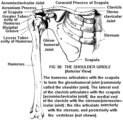
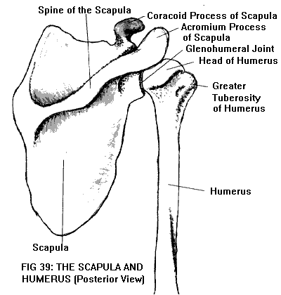
The upper end or head of the humerus is shaped like a hemisphere (actually, somewhat less than half a sphere). Adjacent to the humeral head are two bony prominences, the greater and lesser tuberosities. Between the two tuberosities (on the anterolateral aspect of the humerus) runs the bicipital groove.
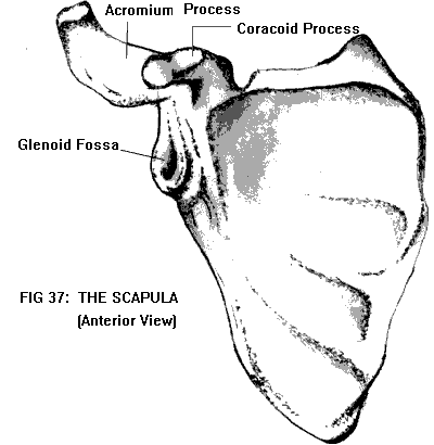
The scapula is a flat, triangular bone that lies over the posterior surface of the rib cage. At its upper lateral corner is a cuplike depression (the glenoid fossa) which forms a socket for the head of the humerus (Figure 37). This glenohumeral joint is the one that is commonly referred to as "the shoulder". The posterior surface of the scapula is divided by a nearly horizontal ridge of bone, the scapular spine. The spine extends laterally to form a projection (the acromion) which overhangs the glenoid fossa. The anterior surface of the scapula, just medial to the glenoid fossa, has a beaklike projection (the coracoid process) that acts as an attachment for muscles and ligaments.
The clavicle is shaped like a rod with a slight S curve; it extends from the base of the neck to the shoulder. Medially it is connected to the upper part of the sternum (breastbone), at the sternoclavicular joint. Laterally it is connected to the acromion of the scapula, at the acromioclavicular joint.
The sternoclavicular joint is the only bony connection between the shoulder girdle and the trunk. The scapula is connected to the trunk indirectly through the acromioclavicular (AC) joint; otherwise, the scapula is attached to the trunk only by muscles.
During scapular movements (e.g., shrugging; elevating the arm overhead), the AC joint permits the clavicle to glide and rotate on the scapula. The AC joint is bound together by the acromioclavicular and coracoclavicular ligaments.
The glenohumeral joint is a ball-and-socket type joint similar to that of the hip (see Anatomy of the Joints). However, unlike the hip, the socket is shallow. This allows great freedom of movement in all directions, but at the cost of stability. The glenoid fossa is deepened somewhat by a fibrous lip or labrum. The labrum is actually a fold of the joint capsule, which encloses the entire joint from the glenoid to the neck of the humerus. The anterior part of the joint capsule is reinforced by several additional folds or thickenings known as the glenohumeral ligaments. Other ligaments are located outside the capsule; these include the coracoacromial and coracohumeral ligaments. The coracoacromial ligament stretches between the acromion and the coracoid process, forming a roof over the head of the humerus, the coracoacromial arch. In addition to protecting and stabilizing the joint, it plays an important role in the mechanics of shoulder movement (discussed below).
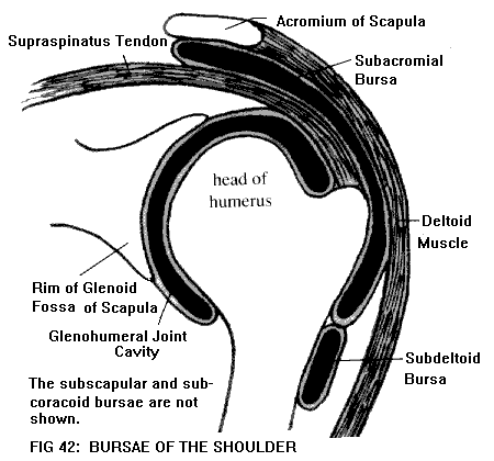
There are numerous bursae in the region of the shoulder (Figure 42). The bursae most commonly involved in pathology are the subacromial bursa (located between the acromion and the joint capsule) and subdeltoid bursa (between the deltoid muscle and the joint capsule). Other large bursae include the subscapular and subcoracoid bursae. The subscapular bursa usually communicates with the joint cavity; the subcoracoid bursa sometimes does so. The subacromial and subdeltoid bursae do not communicate with the joint cavity, but often connect with each other (Figures 40 & 41).
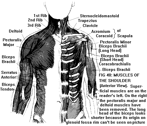
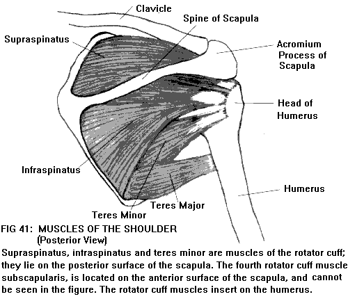
Movement at the glenohumeral joint can take place in all directions: flexion (raising the arm in front) and extension (raising the arm in back); abduction (raising the arm outward to the side) and adduction (bringing the arm in to the side); internal and external rotation; and circumduction (a combination of all of these). Each movement is brought about by different groups of muscles.
The deltoid is the muscle that covers the point of the shoulder. It originates on the clavicle and on the scapular spine and acromion; it inserts into the lateral side of the humerus. The deltoid abducts the glenohumeral joint; however, by itself it can abduct the humerus to an angle of only 90o (relative to the scapula). At that point, the greater tuberosity of the humerus comes up against the coracoacromial arch. In order to abduct further, the head of the humerus must be externally rotated so that the tuberosity is turned out of the way.
Rotation of the humerus is accomplished by a group of four muscles, subscapularis, supraspinatus, infraspinatus, and teres minor, collectively called the rotator cuff. These muscles originate on different parts of the scapula, and insert like a cuff around the perimeter of the humeral head, where their tendons blend with the joint capsule. In addition to externally and internally rotating the humerus, the rotator cuff helps stabilize the joint during abduction by pulling the humeral head into the glenoid fossa.
With external rotation, the glenohumeral joint can be abducted to an angle of about 120o before the humeral head once more impinges on the coracoacromial arch. Further elevation of the arm requires rotation of the scapula (counterclockwise as viewed from behind, so that the glenoid fossa tilts upward). This provides an additional 60o of elevation, for a total of 180o (arm straight overhead). Scapular rotation is accomplished by the trapezius and serratus anterior muscles. (Trapezius originates on the cervical and thoracic spine, and inserts on the scapula. Serratus anterior originates on the anterior rib cage and inserts on the scapula.)
During the first 15-20o of abduction, movement takes place almost exclusively at the glenohumeral joint. With further abduction, the scapula comes into play; both glenohumeral and scapular movement then take place simultaneously. Since the scapula and clavicle are connected, when the scapula rotates the clavicle also moves, at both the sternoclavicular and acromioclavicular joints. Muscles that flex the glenohumeral joint include pectoralis major (originating on the clavicle, sternum, and rib cartilages; inserting on the humerus) and coracobrachialis (originating on the coracoid process; inserting on the humerus). Muscles that extend the humerus include latissimus dorsi (originating in the thoracolumbar region and on the pelvis; inserting on the humerus) and teres major (originating on the scapula; inserting on the humerus). Both pectoralis major and latissimus dorsi also adduct and internally rotate the humerus.
Another muscle that is anatomically related to the shoulder is biceps brachii. Biceps has two parts, a "long head" and a "short head". The short head originates on the coracoid process; the long head, on the upper margin of the glenoid fossa. The tendon of the long head passes through the capsule of the glenohumeral joint, and emerges in the bicipital groove between the greater and lesser tuberosities of the humerus. Encased in a synovial sheath, the tendon glides smoothly in the bicipital groove during movements of the humerus; it is kept from slipping out of the groove by the transverse humeral ligament. The two heads of biceps come together distally and insert on the radius, just below the elbow. Although its tendon passes through the shoulder, biceps is not primarily a mover of the shoulder. Its main actions are supination of the forearm (rotation of the forearm palm upward) and flexion of the elbow.

