|
|
||||||||||||
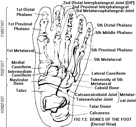
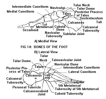
FOOT AND ANKLE (see also Anatomy of the Joints)The foot is considered to have three subdivisions: the forefoot (front part of foot, including toes), midfoot, and hindfoot (rear part of foot, including heel). The midfoot and hindfoot together are sometimes referred to as the tarsus, because they are composed of seven tarsal bones. The tarsal bones are irregular in shape and size; their interlocking shapes enable them to form a highly stable arrangement (the arches of the foot). The ankle is the joint between the tarsus and the lower leg. The bones of the forefoot, midfoot, and hindfoot are shown in Figures 13 & 14.
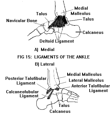
Forefoot:The framework of the forefoot is formed by five metatarsal bones, along with the phalanges (the bones of the digits or toes). Each digit has three phalanges (proximal, middle, and distal), except for the big toe, which has two (proximal and distal). The digits and their metatarsal rays are numbered from one to five, starting with the big toe. The metatarsals and phalanges are long bones. Each has a diaphysis (shaft) with slightly flaring ends. The proximal end or base of each bone has a smooth articular surface where it forms a joint with the adjacent bone. The distal end or head also has an articular surface, except for the distal phalanges, whose distal ends provide attachment for the soft tissue (pulp) of the digit tips. Of the metatarsal bones, the first bears the most weight and plays the most important role in propulsion; it is therefore the shortest and thickest. It provides attachment for several tendons, including tibialis anterior and peroneus longus (see below, under Muscles and tendons). The fifth metatarsal has a tuberosity (protuberance) on the lateral side of its base, to which the peroneus brevis tendon is attached. (The tuberosity of the fifth metatarsal can be felt halfway along the lateral side of the foot.) The second, third, and fourth (called the internal metatarsals) are the most stable of the metatarsals, in part because of their protected position; but also because they have only minor tendon attachments, and therefore are not subjected to strong pulling forces. The joints between the metatarsals and the proximal phalanges are called the metatarsophalangeal (MTP) joints. Each digit also has two interphalangeal (IP) joints, proximal (PIP) and distal (DIP), except for the big toe, which has only one IP joint. Each MTP and IP joint is bound together by several ligaments,one on each side of the joint (medial and lateral collateral ligaments), and one along the plantar (sole) surface (plantar ligament). The first MTP joint has an additional feature. Near the head of the first metatarsal, on the plantar surface of the foot, are two sesamoid bones. (A sesamoid is a small, oval-shaped bone which develops inside a tendon, where the tendon passes over a bony prominence. In this case, the tendon is that of flexor hallucis brevis, as it passes over the first metatarsal head.) These sesamoid bones articulate with the head of the first metatarsal, and function as part of the first MTP joint. They are held in place by their tendons, and are also supported by ligaments. These include the sesamoid collateral ligaments (which bind the sesamoids to the metatarsal head) and the intersesamoidal ligament (which connects the sesamoids to each other). Midfoot:The midfoot is composed of five of the seven tarsal bones, the navicular, cuboid, and three cuneiform bones. These can be thought of as being arranged in two irregular rows, with the cuboid occupying space in both rows. The proximal row contains the navicular (on the medial side of the foot) and the cuboid (on the lateral side). The distal row contains the three cuneiforms (medial, intermediate, and lateral) and the cuboid (lateral to the lateral cuneiform). The boundary between the midfoot and forefoot consists of five tarsometatarsal (TMT) joints, the joints between the distal row of the midfoot and the bases of the metatarsals. (The medial, intermediate, and lateral cuneiforms articulate with the first, second, and third metatarsals, respectively; the cuboid articulates with the fourth and fifth metatarsals.) There are also multiple joints within the midfoot itself. The distal row of the midfoot has two intercuneiform joints (between adjacent cuneiforms) and a cuneocuboid joint (between the lateral cuneiform and the cuboid). Proximally, the three cuneiforms articulate with the navicular bone (the cuneonavicular joints). In some individuals, there is also a small articulation between the cuboid and navicular. In addition to its articular surfaces, each tarsal bone has specific features adapted for function. For example, the medial surface of the navicular projects downward to form a tuberosity, which serves as an attachment for the tibialis posterior tendon. The lateral surface of the cuboid also has a tuberosity, which serves as a ligament attachment. The cuboid bone has no major tendon attachments; however, the peroneus longus tendon passes across the cuboid tuberosity, to run in a groove on the plantar surface of the bone. The peroneus longus tendon often contains a sesamoid bone, which articulates with a small facet (articular surface) on the tuberosity. Hindfoot:The remaining two tarsal bones, the talus (also called astragalus) and calcaneus, make up the hindfoot. Calcaneus is the largest tarsal bone, and forms the heel. The talus rests on top of it, and forms the pivot of the ankle. The shape of calcaneus is complex. On its upper surface are three smooth facets, posterior, middle, and anterior, which articulate with corresponding facets on the lower surface of the talus to form the subtalar joint (Figure 12). Of these three talocalcaneal facets, the posterior is the largest, covering almost the entire width of the calcaneal body. The middle and anterior facets are located on the medial side of the upper calcaneal surface, and are usually continuous with each other. The middle and posterior facets are separated from each other by a deep groove, which together with a corresponding groove on the talus, forms a channel between the two bones called the sinus tarsi. The lateral wall of calcaneus is nearly flat, except for a small ridge called the peroneal tubercle. The medial wall has a shelf-like projection, the sustentaculum tali. This shelf carries the middle talocalcaneal facet on its upper surface. The undersurface of the shelf has a groove for the flexor hallucis longus tendon. The front or anterior process of calcaneus articulates with the cuboid bone to form the calcaneocuboid joint. The rear part of calcaneus consists of a large rounded projection, the calcaneal tuberosity, which forms the back of the heel and provides attachment for the Achilles tendon. The undersurface of the tuberosity forms the bottom of the heel; this surface comes into contact with the ground during weightbearing, cushioned by a fibroelastic fat pad. The talus, which rests on top of calcaneus, also has a complex shape. The main part or body of the talus is roughly cubical. Its smooth, dome-shaped upper surface (the talar dome) articulates with the distal ends of the tibia and fibula (the two bones of the lower leg) to form the ankle joint (see The ankle, below). The rear surface of the talar body protrudes backward to form a posterior process. In some individuals, the posterior process ossifies (develops into bone) independently, and may remain separate from the talar body as a small accessory bone called the os trigonum (see also Ossicles, below). Projecting forward from the talar body is the head of the talus, which is separated from the body by a slight constriction called the neck. The talar head articulates with the navicular bone, forming the talonavicular joint. The lower surface of the talus contains three smooth facets, posterior, middle, and anterior, which articulate with the corresponding facets on the upper surface of calcaneus. The posterior facet (the largest of the three) covers the undersurface of the talar body; the anterior and middle facets are located on the undersurface of the talar head. Together, these three talocalcaneal articulations form the subtalar joint. The subtalar joint is bound together by several talocalcaneal ligaments. In addition, it is supported by portions of the medial and lateral ligaments of the ankle, which span both the ankle and the subtalar joint (see The ankle, below). The calcaneocuboid and talonavicular joints, together referred to as the midtarsal joint, form the boundary between hindfoot and midfoot. Ossicles:The foot contains a variable number of ossicles or small bones. These are of two types, sesamoid bones and accessory bones. Sesamoids are small bones that develop inside a tendon, where the tendon passes over a bony prominence. The two sesamoid bones of the first MTP joint have already been mentioned; these are a constant feature. The other MTP joints only occasionally have sesamoid bones. The peroneus longus tendon frequently contains a sesamoid bone, at the point where it passes over the cuboid tuberosity. Sesamoids may also occur in other locations in the foot. The foot may also contain ossicles that are not associated with a tendon, but result from developmental variations. In the fetus, the skeleton initially consists of cartilage, which gradually ossifies (turns to bone) during fetal development and childhood. Each bone has a primary ossification center. The process of ossification progresses outward from this center, until the bone is completely ossified. In long bones, the primary center is located in the middle of the shaft; later, secondary ossification centers develop at the ends of the long bone. Irregularly shaped bones such as the tarsal bones may also develop secondary centers. In some individuals, complete ossification does not occur; the secondary center remains separate from the rest of the bone, forming an accessory ossicle. An example is the os trigonum (mentioned earlier), which arises as a secondary ossification center in the posterior process of the talus. About 50% of individuals have an os trigonum. Accessory ossicles may also occur in other locations in the foot. Movements of the foot and toes:Toe movements take place at the IP and MTP joints. These joints are capable of motion in two directions: plantar flexion (bending toward the sole of the foot) and dorsiflexion (bending toward the dorsum or top of the foot). In addition, the MTP joints permit abduction (spreading apart) and adduction (bringing together) of the toes. Abduction ordinarily accompanies dorsiflexion of the toes; adduction accompanies plantar flexion. The foot as a whole (excluding the toes) has two movements: inversion (turning the sole inward) and eversion (turning the sole outward). All the joints of the hindfoot and midfoot, from the subtalar to the TMTs, contribute to these movements, which are complex and consist of several components. Inversion includes components of medial rotation (toeing inward) and supination (rotating the medial border of the foot upward). Eversion includes components of lateral rotation (toeing outward) and pronation (rotating the lateral border of the foot upward). In addition, foot movements ordinarily are combined with ankle movements (see The ankle, below). The ankle:The ankle is the joint between the foot and the leg, the articulation of the talus with the distal tibia and fibula (Figures 16 & 17). The distal end of the fibula forms the lateral malleolus, the bony protuberance that can be seen and felt on the lateral side of the ankle. The distal end of the tibia has a smooth concave articular surface, the tibial plafond, as well as a bony projection that forms the medial malleolus. (The posterior margin of the distal tibia is sometimes referred to as the posterior malleolus. However, unlike the medial and lateral malleoli, it is not a prominent landmark.) Together, the distal tibia and fibula form a mortise, a rectangular recess into which the talus fits. The tibial plafond articulates with the talar dome. The medial and lateral malleoli articulate with facets on the medial and lateral sides of the talar body, respectively. (These talar facets are continuous with the articular surface of the dome.) These articulations allow the talus to pivot within the mortise. The ankle joint is capable of two movements, dorsiflexion and plantar flexion. These ankle movements are ordinarily accompanied by eversion and inversion of the foot, respectively. The ankle is supported by two groups of ligaments, medial and lateral (Figure 15). The medial ligaments bind the medial malleolus to the talus, calcaneus, and navicular bone; together, they are known as the deltoid ligament. The lateral ligaments bind the lateral malleolus to the talus and calcaneus. There are three lateral ligaments: two talofibular ligaments (anterior and posterior) and one calcaneofibular ligament. The calcaneofibular ligament and the tibiocalcaneal portion of the deltoid ligament span not only the ankle but also the subtalar joint. The tibionavicular portion of the deltoid ligament spans both the ankle and the talonavicular joint.
Just above the ankle mortise, the distal tibia and fibula articulate with each other to form the distal tibiofibular syndesmosis, a fibrous joint that permits only slight motion. The syndesmosis is bound together by the distal tibiofibular ligaments (anterior and posterior). Along with the interosseous membrane (which connects the shafts of the tibia and fibula), these ligaments maintain the stability of the mortise. The arches of the foot:The foot has two important functions: weightbearing and propulsion. These functions require a high degree of stability. In addition, the foot must be pliable, so it can adapt to standing and walking on uneven surfaces. The multiple bones and joints of the foot give it pliability; however, such a segmented structure cannot bear weight unless the segments are arranged in the form of an arch. The foot may be considered to have three arches. The medial longitudinal arch (of the medial border of the foot) is the highest and most important of the three arches (Figure 19A). It is composed of the calcaneus, talus, navicular, cuneiforms, and the first three metatarsals. The talus occupies the highest point of the arch; with its head wedged between the calcaneus and navicular, it is the "keystone" that holds the arch together. The lateral longitudinal arch (of the lateral border of the foot) is lower and flatter than the medial arch (Figure 19B). It is composed of the calcaneus, cuboid, and the fourth and fifth metatarsals, with the cuboid as the keystone. The transverse arch (at right angles to the longitudinal arches) is composed of the cuneiforms, the cuboid, and the five metatarsal bases. The wedge shapes of the cuneiforms help hold the transverse arch together. The arches of the foot are maintained not only by the shapes of the bones themselves, but also by the ligaments that tie the bones together. In addition, muscles and tendons play an important role in supporting the arches (see under Muscles and tendons, below). Muscles and tendons:The muscles of the foot are classified as either intrinsic or extrinsic. The intrinsic muscles, so called because they are located within the foot itself, are responsible for movements of the toes. They include flexors (plantar flexors), extensors (dorsiflexors), abductors, and adductors of the toes. (Flexor hallucis brevis, whose tendon contains the sesamoid bones of the first MTP joint, is an intrinsic plantar flexor of the big toe.) Various intrinsic muscles also help support the arches of the foot. The extrinsic muscles are so called because they are located outside the foot, in the lower leg. They have long tendons that cross the ankle, to insert (attach) on the bones of the foot, except the talus, which has no tendon attachments (Figure 18). Altogether there are thirteen tendons that cross the ankle. They are responsible for movements of the ankle, foot, and toes; some of these tendons also help support the arches of the foot. The extrinsic muscles and their tendons are described below.
Gastrocnemius and soleus, located in the posterior calf, are the main plantar flexors of the ankle. They unite to form a common tendon, the calcaneal or Achilles tendon, which descends vertically over the back of the ankle to insert on the calcaneal tuberosity. Tibialis posterior, located deep inside the calf, is the main invertor of the foot; in addition, it assists gastrocnemius and soleus with plantar flexion of the ankle. Its tendon descends on the medial side of the ankle, passes behind and beneath the medial malleolus, and continues onto the foot. It has multiple insertions, including the navicular tuberosity. Tibialis anterior, located in the front of the leg, is the main dorsiflexor of the ankle; in addition, it assists tibialis posterior with foot inversion. Its tendon descends over the front of the ankle, passing underneath a band of fibrous tissue called the extensor retinaculum, which holds the tendon in place. The tendon continues over the dorsum onto the medial side of the foot, to insert on the medial cuneiform and first metatarsal. Peroneus brevis and peroneus longus, located on the lateral side of the leg, are the main evertors of the foot; in addition, they assist with plantar flexion of the ankle. Their tendons descend together on the lateral side of the ankle, passing behind and beneath the lateral malleolus. The tendons continue forward along the lateral surface of the calcaneal body (brevis passes above the peroneal tubercle, longus below). Peroneus brevis then inserts on the tuberosity of the fifth metatarsal. Peroneus longus crosses the cuboid tuberosity, runs in a groove on the underside of the cuboid bone, and continues across the sole of the foot to insert on the first metatarsal and medial cuneiform. Flexor hallucis longus, located deep inside the calf, is primarily a plantar flexor of the big toe; however, it also assists with plantar flexion of the ankle and inversion of the foot. Its tendon descends on the medial side of the ankle, alongside the tibialis posterior tendon. After passing beneath the medial malleolus, the tendon continues onto the sole of the foot, and inserts on the distal phalanx of the big toe. Flexor digitorum longus, located deep inside the calf, is primarily a plantar flexor of the second through fifth toes; however, like flexor hallucis longus, it also assists with plantar flexion of the ankle and inversion of the foot. Its tendon descends on the medial side of the ankle, alongside tibialis posterior and flexor hallucis longus. After passing beneath the medial malleolus, the tendon continues onto the sole of the foot, where it separates into four divisions. The four divisions insert on the distal phalanges of the second through fifth toes. Extensor hallucis longus, located in the front of the leg, is primarily a dorsiflexor of the big toe; however, it also assists with ankle dorsiflexion and foot inversion. Its tendon descends over the front of the ankle, passes under the extensor retinaculum, and continues onto the dorsum of the foot, to insert on the distal phalanx of the big toe. Extensor digitorum longus, located in the front of the leg, is primarily a dorsiflexor of the second through fifth toes; however, it also assists with ankle dorsiflexion and foot eversion. It gives rise to five tendons, which descend together over the front of the ankle and pass under the extensor retinaculum. As they continue onto the dorsum of the foot, the tendons diverge. Four of them insert on the distal phalanges of the second through fifth toes. The fifth, called peroneus tertius, inserts on the fifth metatarsal. (Peroneus tertius does not contribute to toe dorsiflexion; however, it helps dorsiflex the ankle and assists with foot eversion.) Except for the Achilles tendon (which descends vertically to insert on the calcaneus), all of the above tendons change their direction from vertical to horizontal as they cross the ankle. The lateral tendons (peroneus brevis and longus) change direction by passing under the lateral malleolus, which acts as a pulley. (Peroneus longus then changes direction a second time, with the cuboid bone as a pulley.) Similarly, the medial tendons (tibialis posterior, flexor digitorum longus, and flexor hallucis longus) change direction as they pass under the medial malleolus. The anterior tendons (tibialis anterior, extensor digitorum longus, and extensor hallucis longus) change directions as they pass onto the dorsum of the foot, with the extensor retinaculum as the pulley. Each tendon, as it passes over its pulley, is enveloped by a tendon sheath lined with synovium. The synovial lining secretes a lubricating fluid that minimizes friction. Only the Achilles tendon has no pulley and no synovial sheath. In addition to producing movements of the ankle, foot, and toes, the tendons of several extrinsic muscles help maintain the arches of the foot. The medial longitudinal arch is supported by tibialis posterior and anterior, as well as flexor hallucis longus and flexor digitorum longus. (Of these, tibialis posterior is the most important.) The lateral longitudinal arch is supported by peroneus brevis and longus, along with the lateral divisions of flexor digitorum longus. The transverse arch is supported by peroneus longus.
|

_______________ Stop Letting Elbow Pain Limit YOU!….. Your
Risk Free Opportunity to Become Pain Free…… ______________ No More TMJD _____________
_______________ Scientifically Proven Shin Splints Treatment |


