|
|
||||||||||||
|
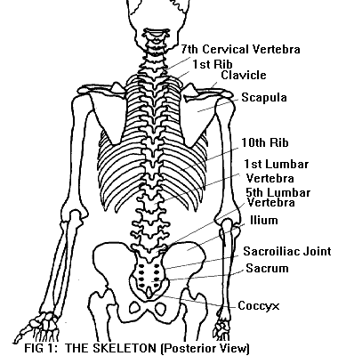
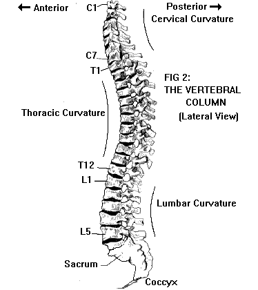
Thanks to Darryl Hosford credit for his work. BACK AND NECKVertebral column:The adult spine consists of 26 bones called vertebrae, arranged in a column (Figures 1 & 2). (In childhood there are 33-35 individual bones, several of which later fuse together.) These bones are named and numbered from the top downward: seven cervical vertebrae (C1-C7) in the neck, twelve thoracic vertebrae (T1-T12) in the upper/middle back, and five lumbar vertebrae (L1-L5) in the small of the back. L5 rests on top of the sacrum, a large triangular bone made up of five fused vertebral segments. Below the sacrum is the coccyx (tailbone), a small bone made up of three to five fused segments. Although the coccyx is at the bottom of the vertebral column, it does not bear weight. The weight of the body is transmitted through the sacrum to the ilium (pelvic bone). The joints that connect the sacrum and the ilium are called the sacroiliac joints.
Viewed from the side, the adult spine has three normal curves (Figure 2). In the cervical and lumbar regions, the convexity of the curve is forward (lordosis); in the thoracic region, the convexity is rearward (kyphosis). The cervical and lumbar lordoses are not present in a newborn infant. The cervical lordosis develops as the infant begins to hold its head up; the lumbar lordosis develops as the child learns to stand erect.
Vertebrae:Although vertebrae of different levels have some structural differences, all have basic features in common (Figure 3). Each vertebra (except C1) has a drum-shaped body which bears most of the weight. Projecting backward from the vertebral body are two short pedicles. Extending back from the pedicles are two bony plates called laminae, which meet in the middle and are fused together. The pedicles and laminae together form a bony arch enclosing a space called the vertebral foramen. (Foramen is Latin for "hole"; the plural is foramina.) With the vertebrae arranged in a column, the vertebral foramina line up to form a hollow tube, the spinal canal. Contained within this canal, surrounded and protected by bone, is the spinal cord.
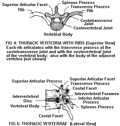
Several other bony projections or "processes" arise from the vertebral arches. The spinous process extends straight backward from the point where the two laminae meet. (The spinous process of C7 or T1 is especially prominent, and can easily be felt at the nape of the neck.) Two transverse processes (one on each side of the vertebra) extend laterally from the junctions of pedicle and lamina. The spinous and transverse processes serve as attachments for muscles and ligaments. Each vertebra also has two superior (upper) and two inferior (lower) articular processes, each with an articular (joint) surface called a facet. The inferior facets of each vertebra articulate with the superior facets of the vertebra below, and so on down the column. These interlocking facet joints stabilize the spinal column, helping to hold it in alignment while permitting flexibility. The facet joints are synovial joints (see also Anatomy of the Joints). In the thoracic region of the spine, the vertebrae also have costal facets which articulate with the ribs (Figure 4). This gives the thoracic spine more structural stability but less flexibility than the cervical and lumbar regions.
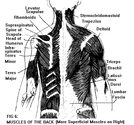
The cervical spine also has some special features. The C1 and C2 vertebrae are unique, and this is reflected in the fact that they are the only individual vertebrae that have names. C1, the atlas, has no vertebral body and no spinous process. It consists of a ring of bone, broadened on each side where it bears the weight of the skull. The joints between the atlas and the base of the skull (the atlanto-occipital joints) allow the head to nod forward or tilt back. C2, the axis, has an odontoid ("toothlike") process that projects upward from the vertebral body. The odontoid process is encircled by the bony ring of the atlas. Held in place by strong ligaments, it acts as a pivot for the atlas. The joints between the axis and atlas (the atlanto-axial joints) allow rotation of the head on the neck. C3-C7 have a more typical structure. However, all seven cervical vertebrae have a feature that is not shared by other vertebrae: a foramen in each transverse process. The two vertebral arteries pass through these foramina (except those of C7) on their way to the brain. Intervertebral discs:Interposed between each pair of vertebral bodies are pads of fibrocartilage called intervertebral discs. The outer part of the disc forms a thick fibrous ring (the annulus fibrosus) which surrounds and contains a soft, gelatinous center (the nucleus pulposus). Between the disc and the adjacent vertebral bodies (above and below) are thin plates of cartilage (the vertebral end plates). The discs give the spine flexibility and act as shock absorbers. Since they are firmly fastened (by the vertebral end plates) to the vertebral bodies, the discs also help hold the spinal column in alignment. Each disc is named and numbered according to the vertebrae above and below it, for example, the C5-6 disc is the disc between C5 and C6; the L5-S1 or lumbosacral disc is the disc between L5 and the sacrum. There is no C1-2 disc, because C1 (the atlas) has no vertebral body. Spinal cord and nerves:In addition to providing support, flexibility, and a site for muscle attachments, the spinal column also houses and protects the spinal cord. The spinal cord is an extension of the brainstem (the lowest part of the brain), extending from the base of the skull down to the low back. Along its length, it gives off 31 pairs of spinal nerves, which branch to form the peripheral nerves of the neck, trunk, and extremities. The origins of the spinal nerves as they emerge from the spinal cord are called nerve roots. The spinal cord is covered by three membranes (the meninges), which are continuous with the membranes that cover the brain. The delicate innermost membrane, called the pia, forms the surface of the spinal cord itself. The weblike middle membrane is called the arachnoid (Greek for "spiderlike"), and the tough outer membrane is the dura. The subarachnoid space (between the arachnoid and pia) is filled with cerebrospinal fluid (CSF), which circulates around the spinal cord and brain. The meninges thus form a fluid-filled sac around the brain and spinal cord, with sleeves for the emerging nerve roots. The spinal nerves exit from the vertebral column through openings between adjacent vertebrae. These openings, called intervertebral foramina, are located just in front of the facet joints. The spinal nerves are named and numbered according to the vertebral levels at which they exit. There are eight paired cervical nerves (C1-C8), twelve thoracic (T1-T12), five lumbar (L1-L5), five sacral (S1-S5), and one coccygeal (Co1). Note that there are eight pairs of cervical nerves, although there are only seven cervical vertebrae. The C1 nerves exit above the C1 vertebra (between C1 and the base of the skull). The C2 nerves exit below the C1 vertebra (between C1 and C2); and so on, down to the C8 nerves, which exit below the C7 vertebra (between C7 and T1). The rest of the spinal nerves exit below the vertebral segment of the same name and number: the T1 nerve below the T1 vertebra; the T12 nerve below the T12 vertebra (between T12 and L1); the L5 nerve below the L5 vertebra (between L5 and the sacrum). The first four pairs of sacral nerves exit through the four pairs of sacral foramina, and so on. Although the spinal nerves correspond to their respective vertebral levels, the spinal cord itself is shorter than the vertebral column, extending only as far as the L1 vertebra. Below that level, the spinal canal contains only the roots of the lumbar, sacral, and coccygeal nerves, as they descend to exit at the appropriate levels. This bundle of descending nerve roots is called the cauda equina (Latin for "horse's tail"). After emerging from the vertebral column, the lower cervical nerves (C5-C8) and the first thoracic nerve (T1) form a network called the brachial plexus, which gives rise to the peripheral nerves of the shoulder and upper extremity. The lumbar and sacral nerves (with a contribution from T12) also form a network, the lumbosacral plexus, which gives rise to the nerves of the lower extremity. The other thoracic nerves (T2-T11) do not form a plexus, but travel independently of each other to supply the trunk. Muscles of the back:(The muscles of the neck are described separately below.) There are two main groups of back muscles, deep and superficial. The deep muscles are responsible for movements of the spine and for maintaining posture. The superficial muscles are responsible for movements of the scapulae (shoulder blades) and shoulders. The deep muscles (sometimes referred to collectively as paraspinal muscles) form a thick mass on each side of the spine, extending from the base of the skull to the sacrum. This muscle mass consists of many separate, overlapping muscles of different lengths, attached to the spinous or transverse processes of different vertebrae. Each individual muscle can be thought of as a string. Pulling a string (contracting a muscle) causes one or more vertebrae to tilt or turn on the vertebra below. Working in coordination, these muscles extend (backward bend) and rotate the spine. (Flexion or forward bending is accomplished by the muscles of the abdomen.) The deep muscles of the back also maintain posture by holding the spine erect against gravity. In the low back, the fascia (fibrous covering) of the deep muscles forms a thick sheet, the lumbar fascia. This fibrous sheet serves as an attachment for other muscles, including muscles of the abdominal wall. Overlying the deep muscles of the back are the superficial muscles (Figure 6). In the upper back, the most superficial of these is trapezius, a large flat muscle that extends like a cape over the upper back and the back of the neck (see also Muscles of the neck, below). Underneath trapezius are the levator scapulae and the rhomboid muscles. Each of these muscles has its origin (attachment of one end) on the cervical and/or thoracic spine, and its insertion (attachment of the other end) on the scapula. Acting together in various combinations, these muscles produce movements of the scapulae (e.g., shrugging the shoulders, bracing the shoulders back).
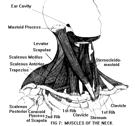
Another group of superficial muscles originates on the scapula and inserts on the upper end of the humerus (upper arm bone). These muscles produce some movements of the upper arm at the shoulder joint (extending the arm backward; raising the arm straight out to the side; rotating the arm inward or outward). Included in this group are supraspinatus, infraspinatus, teres major, and teres minor (all located in the upper back, overlying the scapula), as well as the deltoid (located over the top of the shoulder and upper arm). Latissimus dorsi is a large superficial muscle that covers the middle and low back. Originating on the lower thoracic spine and lumbar fascia, and inserting on the upper end of the humerus, it also is responsible for some shoulder movements. Muscles of the neck:The deep muscles of the neck are a continuation of the deep muscles of the back (described above). They consist of separate, overlapping muscles of different lengths, connecting the cervical vertebrae to each other and to the base of the skull. Attached to the spinous or transverse processes, these muscles extend (backward bend), side bend, and rotate the head and neck. Another group of muscles connects the fronts of the cervical vertebrae to each other and the base of the skull; these muscles flex (forward bend) the head and neck. The superficial muscles of the neck are shown in Figure 7. The scalene muscles are located in the sides of the neck. There are three scalenes on each side: scalenus anterior, medius, and posterior. These muscles originate on the transverse processes of the cervical vertebrae, and insert on the first and second ribs. When the scalenes of one side act alone, they bend and rotate the neck to that side. When both sides act together, they flex the neck slightly.
As mentioned above (in Muscles of the back), the trapezius is a large, flat, superficial muscle that covers the back of the neck and extends like a cape onto the upper back. It originates on the base of the skull and the spine, down to T12; it inserts on the scapulae. The trapezius acts with other muscles to produce scapular movements (shrugging, bracing the shoulders back). However, if the scapula is kept immobile, the trapezius produces movements of the head. Contraction of one side of trapezius tilts the head to that side; contraction of both sides together tilts the head backward. The two sternocleidomastoids (SCMs) are superficial, strap-like muscles located in the front of the neck (one on each side). Each SCM originates at the base of the neck on the sternum (breastbone) and clavicle (collarbone), and inserts behind the ear on the mastoid process of the skull. When one SCM acts alone, it bends the neck to that side, at the same time rotating the head toward the opposite side. When both act together, they flex the neck while extending the head. Underneath the SCMs, in the front of the neck, is a group of strap-like muscles that produce movements of the tongue during swallowing. Other structures of the neck:The neck contains several visceral (internal organ) structures: the upper part of the esophagus, the larynx and upper part of the trachea, and the thyroid gland. The neck also contains major blood vessels (the vertebral arteries, carotid arteries, and jugular veins), as well as numerous lymph nodes.
|
Your
Risk Free Opportunity to Become Pain Free…… ______________ No More TMJD _____________
_______________ Scientifically Proven Shin Splints Treatment _______________
|


